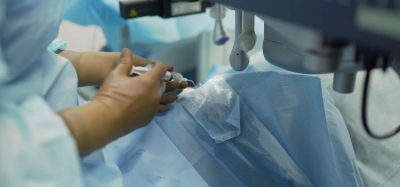
Microbial contamination detection with rapid spectroscopy
The detection method was designed for continuous culture monitoring of cell therapy products and required minimal sample preparation, research showed.
Researchers have demonstrated a novel approach that uses machine-learning aided UV absorbance spectroscopy to detect contamination during the manufacture of cell therapy products.
Based on the findings, the cost-effective and non-invasive method provides a “rapid output (30 minutes) with minimal sample preparation and sample volume (<1mL)”.
In their paper, the researchers stated that considering the limitations of current manufacturing processes, there is a need for sterility testing of cell therapy products to ensure their safety for patient use. Moreover, “with the lack of sterility test frameworks specific to ATMPs, the traditional USP <71> has been the gold standard for sterility testing” of cell therapy products.
A further benefit of the UV absorbance spectroscopy method is that it offers a simpler workflow. There is no need for “extra inoculation, growth enrichment mediums and additional incubation time”, the authors explained.
Results of the machine-learning aided spectroscopy method study
this spectroscopy-based method has value as a preliminary sterility test during different stages of cell product manufacturing [and could bed used] where cultures can be monitored real-time to detect early onset of microbial contamination”
In terms of results, by inoculating “seven bacterial species into mesenchymal stromal cells supernatant aliquots from six commercial donors” contamination events were detected with “mean true positive and negative rates of 92.7 percent and 77.7 percent respectively”, Chelvam et al. noted. They added that specificity improved to 92.0 percent once donor variation was accounted for.
The method facilitated comparable sensitivity to compendial USP <71> test (~24 hours), the authors wrote. In cells spiked with 10 CFUs/mL of E.coli, contamination was detected at the 21-hour timepoint.
To further reduce the time taken for detection using the method, “integration of automated sampling systems to interface between bioreactors and downstream PATs” could be applied, the researchers suggested.
Future application
Overall, this spectroscopy-based method has value as a preliminary sterility test during different stages of cell product manufacturing, according to Chelvam et al. Specifically, it has merit for use “where cultures can be monitored real-time to detect early onset of microbial contamination”.
This paper is available from Research Square as a preprint.