Tool enables imaging of neural activity with near-infrared light
Posted: 23 January 2019 | Nikki Withers (European Pharmaceutical Review) | No comments yet
NIR-GECO1 could help the development of effective treatments for pressing health conditions, including neurodegenerative diseases


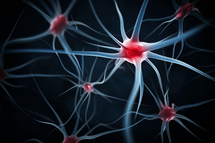
Researchers have developed a tool for visualising neural activity which has the potential to help determine the efficacy of therapeutic drugs at the cellular level.
“This genetically encoded calcium ion (Ca2+) indicator (GECI), designated NIR-GECO1, enables imaging of Ca2+ transients in cultured mammalian cells and brain tissue with sensitivity comparable to that of currently available visible-wavelength GECIs,” write the study authors in Nature Methods.
“Specifically, it emits near-infrared light in the absence of calcium ions. When the concentration of calcium ions increases, it turns dark,” explained Robert Campbell, professor in the Department of Chemistry at the University of Alberta and lead author of the study. “When a neuron ‘fires’ the concentration of calcium ions temporarily increases inside of the cell. We see this as a dimming of the emitted near-infrared light.”
The research builds on previous work in Campbell’s lab focused on developing a toolkit for visualising and manipulating individual neurons. NIR-GECO1 is a protein encoded into DNA, making it most useful for cultured cells in a lab or in model organisms.
“Tissue is relatively transparent to near-infrared light, so this tool has the potential to enable researchers to visualise neuronal activity deeper within the brain than is currently possible,” said Campbell. “This could lead to important insights in the areas of learning and memory, stroke prevention and recovery, and neurodegenerative diseases.”
The researchers conclude: “We demonstrate that NIR-GECO1 opens up new vistas for multicolor Ca2+ imaging in combination with other optogenetic indicators and actuators.”


